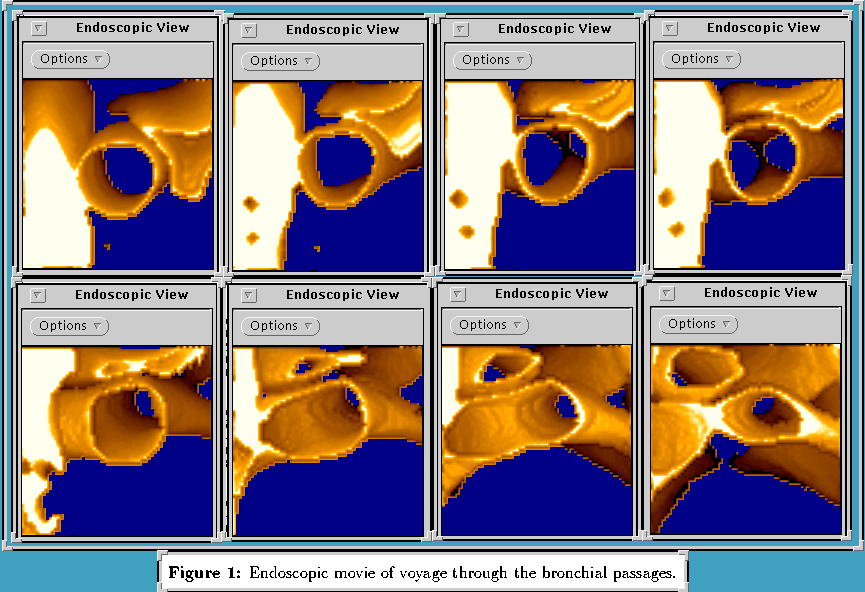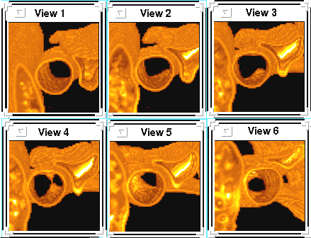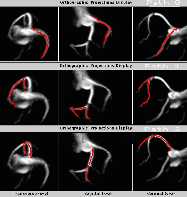
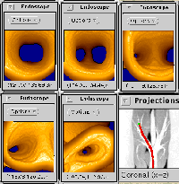
1. Path 1
Path goes through the bronchial passages, starting in the trachea
and heading into the middle lobe of the right lung. Parallel-projection
viewing geometry constructs the rendered views. |
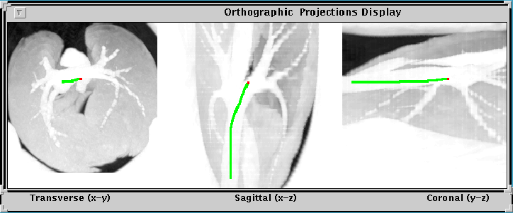
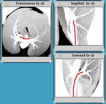
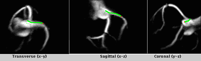
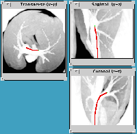
2. Trajectory for Path 1
Path trajectory sketched on supplementary orhtographic projection
displays for voyage seen in picture 1. |
|
|
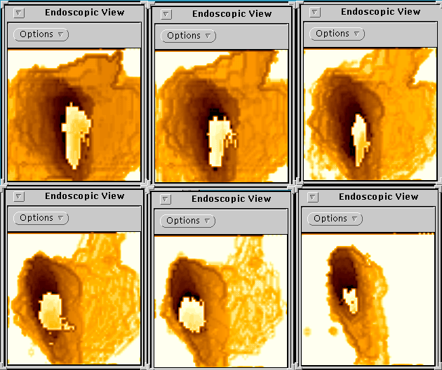
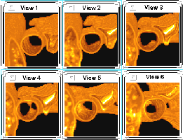
3. Path 2
through the bronchial passages, using the coherence
method for dynamic navigation. The path starts in the trachea
and heads into the upper lobe of the left lung. A perspective-projection
viewing geometry is used in constructing the rendered views. |
4. Path 3
through the bronchial passages, using the ADRT
method for dynamic navigation. The path starts in the trachea
and heads into the middle lobe of the right lung. A parallel-projection
viewing geometry is used in constructing the rendered views. |
|
|
5. Trajectory for Path 3 |
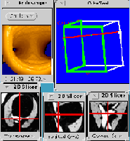
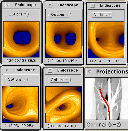
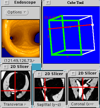
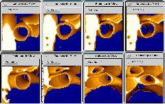
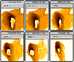
6. Composite view
of various QUICKSEE tools at a site in the right lung's middle
lobe. Endoscope shows the rendered view at the site. 2D Slicer
gives transverse, sagittal, and coronal data slices at the viewing
site. |
|
|
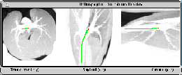
7. 3D voyage through the coronary arteries |
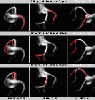
8. Various paths through the coronary arteries |
|
|
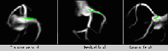
9. Trajectory for above voyage
Path trajectory sketched on supplementary orthographic projection
displays. |